
How to improve your margins and digital dental scans
By Sarfraz Ahmed
Excellent margins are vital for attaining a well-becoming healing requiring minimum chair-facet adjustments. It is essential to make certain the margins are designed and prepped correctly, and afterward, it’s miles first-class to shine your prep. Your preference of margin training relies upon at the kind of crown decided on for the affected person. The forms of margins and whilst to apply them are mentioned beneath.

Knife-part or feather-part margins are appropriate for:
Less teeth discount is needed with a knife-part or feather-part margin, however, this training is much less perfect for layered Zirconia restorations and can purpose becoming issues.

Bevel-chamfer margins are appropriate for:
PFMs.
Bevel-chamfer margins provide a higher marginal in shape, and there may be much less risk a darkish line may be seen on the margin. This margin training isn’t an premiere preference for layered zirconia or IPS e.max crowns.

Chamfer margins are appropriate for:
Chamfer margins offer a greater passive and higher marginal in shape without a darkish margins. They are much less appropriate for layered zirconia and IPS e.max restorations.
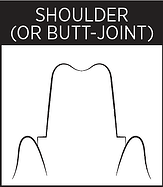
Shoulder or butt-joint margins are appropriate for:
Shoulder or butt-joint margins offer a greater passive in shape and put off darkish margins. This training is suggested for LiS2 and layered zirconia. Care ought to be taken to make certain right isolation of the margin all through training.

Shoulder-bevel margins are appropriate for:
Full zirconia crowns.
Shoulder-bevel margins assist make certain a greater passive and right marginal in shape, getting rid of darkish margins. They aren’t a premiere preference for layered Zirconia crowns, and right margin isolation is vital all through training.
Isolating the margins is vital for a well-becoming crown. If your dental lab can not see the margins of a traditional influence or if a virtual scanner fails to choose up the margins satisfactorily, there may be a excessive hazard the case may be delayed, in particular if the influence is rejected. Otherwise, if the case is going ahead, it may bring about open or quick margins. For those reasons, increasingly more clinicians are selecting to apply virtual impressions.
Digital generation permits records to be captured and visualized, offering the possibility to investigate tiny information which aren’t effortlessly seen on traditional tray impressions.
How to Get a Great Margin Scan on Your iOS
Although virtual impressions can offer precise images, there are numerous critical elements to consider.
Obtaining a Sub-Marginal Line of Sight with Accuracy
The picture displayed withinside the replicate is the picture so that it will be captured with the aid of using the scanner, and ideally, it have to display 0.5mm sub-marginally. Dental labs regularly obtain virtual impressions in which moisture and tissue had been inadequately controlled. Although the margin is seen, it isn’t truly defined. Keep in thoughts that virtual scanners best seize the picture this is seen; the conventional influence method can bodily pressure a few tender tissue out of the manner, as low viscosity fabric is injected into the sulcus. Your techniques, whilst making ready for a virtual influence, ought to will let you visualize the sub-marginal line of sight truly.
Retracting Soft Tissue
Retracting tender tissue is first-class completed with the aid of using packing a wire into the sulcus earlier than taking the influence, however this assignment is frequently less difficult stated than done. Inflamed gingival will regularly bleed, and setting apart the margins is difficult whilst area is limited. Supragingival margins provide a clearer visualization, however frequently enamel which might be being prepped for a crown can have formerly been restored with sub-gingival margins, and the fitness of the gingival tissues is regularly compromised.
Usually, as a minimum a part of the training is beneath the loose gingival margin. If your teeth training has subgingival margins, the tissue ought to be separated from the training part, permitting an awesome best virtual test. A excessive-readability test is completed with top notch best retraction, revealing a clean margin.
Exposing Margins with a Soft Tissue Laser and Retraction Paste
Using a tender tissue laser may be beneficial for exposing sub-gingival margins and for getting rid of dangerous and infected gingival tissue that can be masking the margin or growing immoderate bleeding. Removing infected tissue additionally allows to stimulate the regrowth of more healthy tissues. After treating regions with a dental laser, peroxide answer eliminates tissue debris. More fairly infected regions may be handled with a retraction paste that allows to offer hemostasis and bodily separation among the margin and tissue. The paste is injected into the sulcus, and a company cotton roll is located over the paste. The affected person bites down at the cotton for among and 5 mins, after which period the paste is rinsed away leaving a clean view of the margin.
Using a Retraction Cord
A retraction wire movements gingival tissue out of the manner of the margin arrangements and is inserted lightly beneath the gum line and into the gingival sulcus across the teeth. Using a retraction wire can displace gum tissue effectively, but it’d additionally purpose bleeding. This bleeding can be complicated to control, making it awkward to seize the margins truly. Some forms of wire are already soaked in an answer of hemodent or may be soaked with the aid of using the clinician earlier than placement.
Using the Double Cord Method
It’s additionally feasible to apply the double wire method; the usage of a double wire can beautify tissue displacement, and margins are higher defined. One trouble with the usage of a unmarried wire is that frequently margin regions are extraordinarily skinny and impressions can greater without problems deform. If the affected person has an in particular deep sulcus, the usage of cords prevents tissue from collapsing over the pinnacle of the wire. When packing the cords, % a small wire at once beneath the margin of the teeth, and % a bigger wire at once on pinnacle of the small wire. The huge wire is left in region for as a minimum 5 mins earlier than being removed. You can then determine whether or not to get rid of the small wire earlier than the influence is taken or to go away it in region.
The accurate margin training and tissue control strategies are important to the whole system of fabricating and becoming a top notch restoration, whether or not the use of a traditional or virtual impression. MS Dental Arts Lab has an high-quality chart for enhancing your margins. This chart is to be had for download, and it’s far a brilliant reference tool.

Our licensed and skilled technicians are constantly to be had for case consultations and might help you with case planning. Get in touch now.
No comment yet, add your voice below!